Family Pinnidae
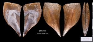
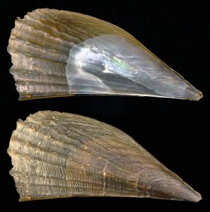
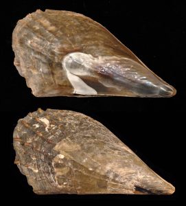
Shell size to 300 mm; shell similar to Atrina rigida, but more delicate. Surface sculpture of about 30 ribs bearing finer, smaller scales. Muscle scar inside nacreous area. Color translucent light-amber to light greenish-brown. This is the most distinctive of the three local species of Atrina. The second image shows an individual in the original National Shell Museum "Live Tank." The bivalve is lying on its side (in nature it would assume a vertical position half-buried in the sand), and the incurrent aperture is on right and excurrent on left. The bivalve can "zip up" the mantle edges to regulate water flow.
Read MoreShell size to 240 mm; shell fan-shaped, triangular. Hinge area straight, representing larger side of triangle. Surface sculpture of narrow ribs separated by larger interspaces; ribs bearing regularly spaced, fluted spines. Ribs present on about top half of fully grown valve. Large muscle scar well within (below) border of nacreous area (pallial line; see photo [middle] of open pen shell by Amy Tripp showing position of muscles). Byssus at pointed extremity anchors penshell into seagrass bottom. Gaping, narrower side of triangle oriented upward. Shell color dark-olive brown. Compare with Atrina rigida (Lightfoot, 1786), which has ribs extending to bottom half of fully grown shells, a muscle scar jutting above the nacreous area, and orange-colored mantle. The additional photos (by José H. Leal) show how the animal can "zip-up" the inner mantle lobe to help regulate the incoming flow of water.
Read MoreShell size to 300 mm; shell fan-shaped, triangular. Hinge area straight, representing larger side of triangle. Surface sculpture of about 15-25 narrow ribs separated by larger interspaces; ribs bearing regularly spaced, fluted spines. Large muscle scar crosses above border of nacreous area (pallial line). Byssus at pointed extremity anchors penshell into seagrass bottom. Gaping, narrower side of triangle oriented upward. Color dark-olive brown. Mantle reddish-orange to brick-orange. The other supplementary image (by Dr. Peter Bush, SUNY/Buffalo) shows the regular arrangement of the platelets (crystals) that comprise the internal, nacreous shell layer. The platelets are very thin, translucent, which imparts the iridescence typical of that kind of shell layer. Compare this species with Atrina seminuda (Lamarck, 1819), which has ribs limited to upper half of fully grown shells, a muscle scar well within the nacreous area, and orange mantle. The small shell (about 57 mm) in the supplementary image on the extreme right was found by Kimberly Nealon on Captiva in December 2015. It illustrates the change in proportions as the shell grows in this genus, with younger individuals being relatively longer. This last image, of a live specimen at low tide, was taken by Amy Tripp near Marco Island.
Read More

